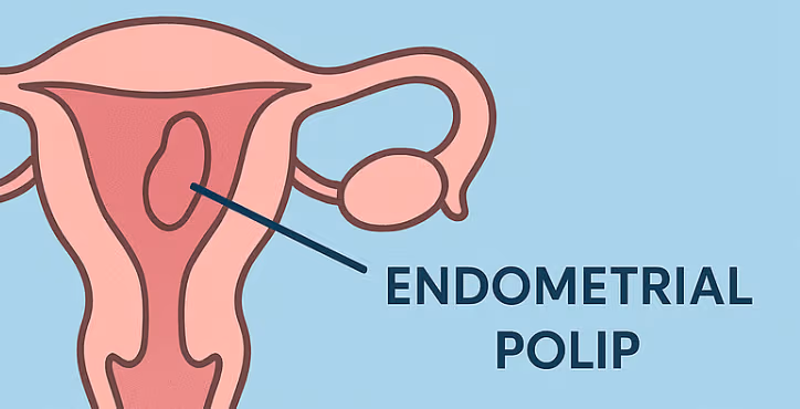
Polyp Surgery

Polyp Surgery

İçindekiler

Endometrial Polyp Surgery: Symptoms, Diagnosis, Treatment and Follow-Up

What is an endometrial polyp?

An endometrial polyp is a localized overgrowth of tissue arising from the inner lining of the uterus, called the endometrium. It projects into the uterine cavity and may be attached by a thin stalk or a broader base. Polyps may be only a few millimeters in size, but some can grow to several centimeters.

Most endometrial polyps are benign. However, because a small proportion may show precancerous or malignant changes, especially in selected risk groups, removed polyps are usually sent for pathological examination.

“Does every endometrial polyp need surgery?”

Not always. Small, asymptomatic polyps in low-risk patients may sometimes be monitored. However, removal is commonly recommended when the patient has abnormal uterine bleeding, postmenopausal bleeding, infertility, recurrent pregnancy loss, suspicious imaging findings, or risk factors for endometrial pathology.

The decision should be individualized according to age, symptoms, menopausal status, fertility goals, polyp size, ultrasound findings and additional risk factors.

What symptoms can endometrial polyps cause?

Some endometrial polyps cause no symptoms and are found incidentally during routine evaluation. When symptoms occur, abnormal uterine bleeding is the most common presentation.

Possible symptoms include:

  • Irregular menstrual bleeding

  • Spotting or bleeding between periods

  • Heavy menstrual bleeding

  • Prolonged periods

  • Bleeding after menopause

  • Spotting after intercourse

  • Difficulty conceiving

  • Association with recurrent pregnancy loss in selected cases

“Is spotting normal, or could it be a sign of a polyp?”

Occasional spotting may be related to hormonal changes, infections, cervical conditions or medications. However, repeated intermenstrual bleeding, bleeding after intercourse or any bleeding after menopause should be evaluated. Endometrial polyps are one possible cause of these symptoms.

Why do endometrial polyps develop?

The exact cause is not always clear. Endometrial polyps are considered estrogen-sensitive lesions, and several factors may increase the likelihood of developing them.

Possible risk factors include:

  • Increasing age

  • Perimenopausal or postmenopausal hormonal changes

  • Obesity

  • Hypertension

  • Tamoxifen use

  • Previous history of endometrial polyps

  • Intrauterine abnormalities detected during infertility evaluation

How are endometrial polyps diagnosed?

The aim of diagnosis is to assess the uterine cavity and the endometrial lining. The choice of test depends on the patient’s symptoms and clinical context.

1. Transvaginal ultrasound

This is often the first-line imaging method. It may show focal thickening, irregularity or a lesion suspicious for a polyp. However, not all polyps are clearly visible on standard ultrasound.

2. Saline infusion sonography

In this method, sterile saline is introduced into the uterine cavity during ultrasound. This helps outline the cavity and can make it easier to distinguish a polyp from other intrauterine lesions such as submucosal fibroids.

3. Hysteroscopy

Hysteroscopy allows direct visualization of the inside of the uterus using a thin camera system. It can be diagnostic and therapeutic at the same time. If a polyp is identified, it can often be removed during the same procedure.

4. Endometrial biopsy

An endometrial biopsy samples tissue from the uterine lining and may be used in the evaluation of abnormal bleeding. However, because a polyp is a focal lesion, blind biopsy may miss it. When an intrauterine structural lesion is suspected, hysteroscopy provides more direct assessment.

“Is hysteroscopy only a diagnostic procedure?”

No. Hysteroscopy can be both diagnostic and operative. The uterine cavity is visualized with a camera, the location and size of the polyp are assessed, and the polyp can be removed with specialized instruments. This is called hysteroscopic polypectomy.

How is endometrial polyp surgery performed?

Endometrial polyp surgery is most commonly performed by hysteroscopic polypectomy. No abdominal incision is required. The surgeon reaches the uterine cavity through the vagina and cervix.

During the procedure:

  1. A thin hysteroscope is gently introduced into the uterus.

  2. The uterine cavity is expanded with fluid to allow visualization.

  3. The polyp is directly identified.

  4. The polyp is removed from its base using appropriate instruments.

  5. The removed tissue is sent for pathological examination.

Hysteroscopic polypectomy is a minimally invasive, uterus-preserving procedure. It is particularly useful in patients who wish to preserve fertility.

Is anesthesia required?

This depends on the setting, the size and number of polyps, patient comfort, additional procedures and the clinician’s preference. Local anesthesia, sedation or general anesthesia may be used. Some small procedures can be performed in an outpatient setting, while others are better suited to an operating room environment.

“Can a polyp come back after removal?”

Yes, endometrial polyps can recur. The recurrence risk varies according to hormonal status, age, associated medical conditions and the structure of the uterine cavity. For this reason, symptom monitoring after treatment is important. If abnormal bleeding returns, the patient should be reassessed.

What is recovery like after surgery?

Most patients recover quickly after hysteroscopic polyp removal. Mild pelvic cramping, light bleeding, spotting or watery discharge may occur for a few days. These are usually temporary.

Medical advice should be sought if any of the following occur:

  • Heavy or persistent bleeding

  • Foul-smelling discharge

  • Fever

  • Severe pelvic or abdominal pain

  • Feeling faint or unwell

  • Unexpected heavy bleeding or possible pregnancy

Patients should follow their doctor’s advice regarding sexual intercourse, tampon use, swimming and strenuous exercise after the procedure.

Why is the pathology result important?

Most endometrial polyps are benign, but microscopic examination confirms the diagnosis and excludes precancerous or malignant changes. Pathology is especially important in patients with postmenopausal bleeding, advanced age or other risk factors. The result may influence further treatment and follow-up.

Why do polyps matter in patients trying to conceive?

In some patients, endometrial polyps may interfere with embryo implantation. They may also be detected during the evaluation of infertility or recurrent pregnancy loss. In patients planning pregnancy, removal may be recommended depending on the size, location and clinical context.

How is follow-up planned?

Follow-up is individualized. In general:

  • The pathology result is reviewed.

  • Improvement in bleeding symptoms is assessed.

  • Fertility plans and timing are discussed when relevant.

  • Any recurrent bleeding, especially after menopause, is reassessed.

  • Follow-up ultrasound or hysteroscopy may be considered when clinically indicated.

When should you see a doctor?

Consult a gynecologist if:

  • You have bleeding between periods

  • Your menstrual bleeding has become significantly heavier

  • You have any bleeding after menopause

  • You are having difficulty conceiving

  • Previous polyp-related symptoms have returned

  • An ultrasound report suggests a possible intrauterine polyp

Conclusion

Endometrial polyps are common intrauterine lesions and are usually benign. Nevertheless, they should be carefully evaluated when associated with abnormal bleeding, infertility or postmenopausal bleeding. Hysteroscopic polypectomy is an effective, minimally invasive and uterus-preserving treatment that allows direct visualization and removal of the polyp. With appropriate diagnosis, treatment and follow-up, most patients achieve good outcomes.

This article is intended for general information only. Diagnosis and treatment decisions should be made by a qualified gynecologist after reviewing the patient’s medical history, examination findings and individual risk factors.